Goals
- Understanding the principle and benefits of magnification.
- Knowing how to use a light microscope.
- Understanding that there are many different kinds of microscopes.
A magnifying glass is a lens that magnifies an object when the glass is held up to it.
A magnifying glass is a double convex lens that is much thicker in the middle than it is at its edges. When light rays pass through the magnifying glass, the double convex lens changes the direction of parallel rays so that they converge and create a virtual image on your eyes' retinas. The more curved the convex lens is, the greater its ability to bend light and magnify images becomes.
Select a small organism, for example a small insect or a part of a plant.
Study the sample by observing it on three levels of magnification:
Write down the things you notice:
Biological organisms and structures come in a large variety of sizes. Some of them can be seen with the naked eye. However, when we use a school microscope or a magnifying glass, we can distinguish structures and organisms we otherwise could not, including cells.
The largest human cell, the ovum, can be distinguished with the naked eye. However, when studying human biology, even smaller things, such as parts of cells, need to be observed. For this reason, medical research makes use of microscopes. Organisms such as pathogens, bacteria and viruses cannot be seen without them.
Although traditional microscopes are useful, even they have limits. Because of this, extremely small structures such as genes must be studied with other methods.
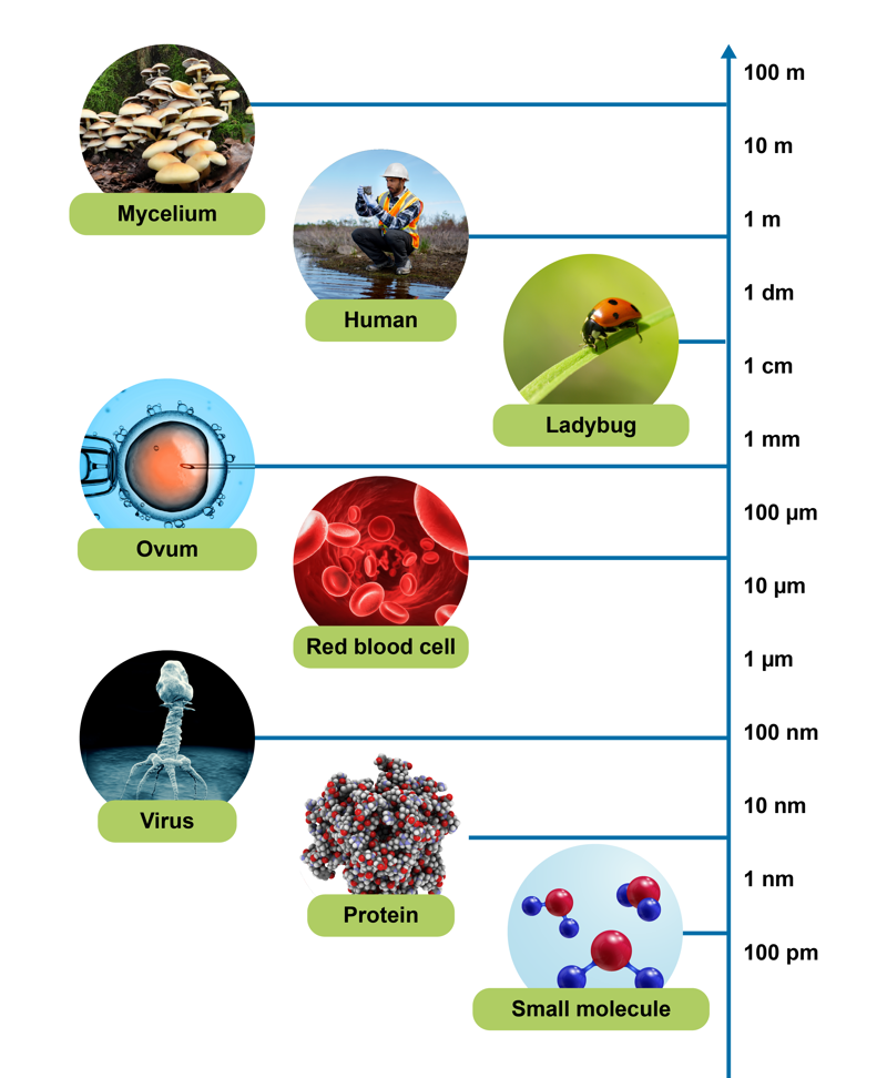
The chart illustrates sizes of various organisms in a scale from one kilometre to one picometre (one trillionth of a metre).
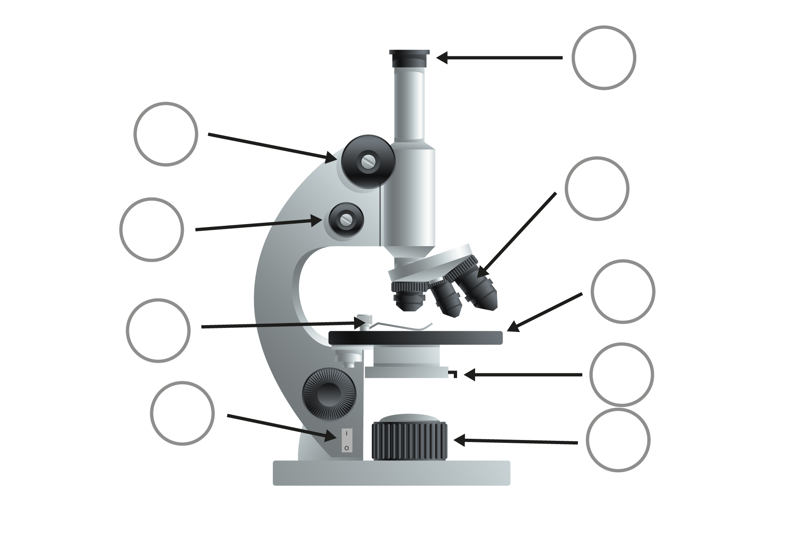
A regular magnifying glass can magnify an image only 15 times. In comparison, the light microscopes used in schools usually magnify the image 40–600 times. Light microscopes accomplish this by using two different kinds of lenses. The eyepiece is called the ocular, and the lenses above the microscope’s stage are called object lenses.
When using a microscope, care must be taken to avoid damage to the device and sample. Make sure your desktop is sturdy enough to avoid vibrations and other movements when looking at small objects. Carry the microscope by holding it from its base and its arm. By making small adjustments, you can make the image clearer while protecting the microscope and the sample.
The Dutch scientist Antonie van Leeuwenhoek was among the first developers of the microscope. Although he lived over 300 years ago, he could already construct precise and functional microscopes.
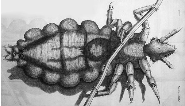
One of the first scientists to make use of van Leeuwenhoek’s invention was the English natural philosopher Robert Hooke, who used a microscope to draw the structure of a head louse (pictured). Hooke was also the first person to use the term cell. He used it to describe the small structures he saw in oak bark.
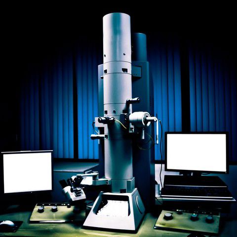
When looking at the fine structure of a cell, even a good light microscope is no longer capable of producing an image that is sufficiently accurate. In this kind of a situation, an electron microscope is required. Electron microscopes provide higher magnifications and higher resolution images, but they cannot be used to study living cells.
The two most important applications of electron microscopy are the Transmission Electron Microscope (TEM) and the Scanning Electron Microscope (SEM). There is a big difference in how these two technologies are used. TEM gives a cross-sectional view of the object, whereas SEM reveals the shape of the object's surface. Thus, TEM is closer to light microscopy in principle. Electron microscopes use an electron beam as their light source. The electron microscope has a much better resolution and depth of field than the light microscope. Electron microscopes, like light microscopes, have a light source and a lens system. The electron beam does not even penetrate the air, let alone the glass lens. Thus, the interior of the electron microscope consists of a vacuum, and the lenses are made of magnets instead of glass.
Transmission electron microscopy, or TEM, is a technique used to observe relatively large structures, from organelles to tissue samples. TEM is also used to study sports injuries, such as tendon and muscle injuries. TEM can be used to view structures that are 100 to 500 nm (nanometres) in size, and it can display details as small as 4 nm. Although electron tomography is a great way to create three-dimensional images of small structures, it is also very difficult. Nowadays, about 100 individual images must be taken and processed. The imaging itself takes two to four hours, whereas the final processing can take an entire day. TEM images are mainly used to examine the cross-sectional structures of cells.
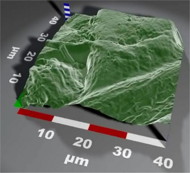
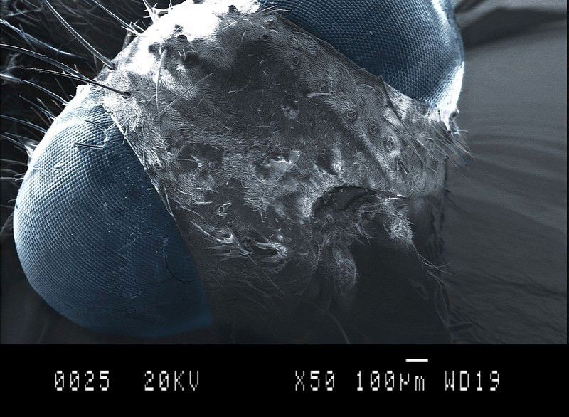
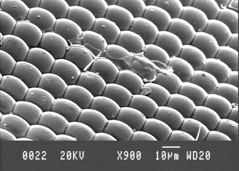
The scanning electron microscope, or SEM, produces images from the surface shape of the sample with a resolution of ten nanometres (10 nm = 10-8 m). SEM images are extremely accurate, and they are easy to understand for those who are used to viewing 3D images. However, SEM cannot be used to create actual three-dimensional images. This would require combining images from different angles.
Because SEM focuses on the composition of the sample’s surface, it is often used to study sample-specific surface reactions. Thus, typical applications of SEM include coating inspections, examining the causes of fractures, and checking surfaces for microbial contamination or rust damage.
The atomic force microscope, or AFM, was developed in the 1980s. Unlike other microscopes, the atomic force microscope is no longer based on visual observation but on "feeling" or "touching" the surface of the sample with a mechanical probe. AFM is a very versatile tool used in electronics and chemistry research. It also has many biological uses.
Units of length include the metre , the centimetre and the millimetre .
One metre is equal to millimetres.
The only human cell we can distinguish without the help of a microscope is the female gamete, the . In comparison, a is too small for the human eye to see.
The resolution of the human eye at a distance of 15 cm is 0.026 mm, or 260 micrometres, at best. We cannot distinguish smaller objects than this. However, we can see very bright objects even if they are even smaller.
The diameter of a red blood cell is about 10 micrometres, whereas the diameter of a bacterium is about 1 micrometre. The diameter of a virus is approximately nanometres. This means that viruses are one hundred times smaller than red blood cells.
Mitä haluat tehdä tekstillä? Teksti käsitellään tekoälyn avulla, eikä sitä sen jälkeen muokata tai tarkisteta. Tekstissä voi esiintyä virheitä. Tarkista tekstin oikeellisuus vertaamalla sitä kirjan alkuperäistekstiin.
Valitse tiedostot, jotka haluat lisätä. Tuetut formaatit ovat txt, html, htm, pdf, odt, odp, ods, xls, xlsx, ppt, pptx, pps, doc, docx, rtf, png, jpg, jpeg ja gif.
| Nimi | |
|---|---|
| poista |
Huom.! Linkkien tulee alkaa ”http://”!
Opiq käyttää verkkosivun toiminnan, turvallisen käytön varmistamisen, käytön analysoimisen ja parhaan käyttömukavuuden tarjoamisen edellyttämiä evästeitä.
Eväste on käyttäjän tietokoneelta verkkosivun palvelimeen lähetettävä pieni tiedosto, joka sisältää verkkosivun toiminnan edellyttämiä käyttäjää ja hänen tekemiä valintoja koskevia tietoja.
Isoin osa evästeistä ovat Opiqin toiminnan kannalta välttämättömiä. Analyyttisistä evästeistä voi luopua ja silloin ei sinun käyttötietojasi ei käytetä Opiqin kehittämiseen. Lue lisää