Individuals consist of cells
All organisms are made up of one or many cells. Most cells are small and invisible to the naked eye. However, every cell contains a large number of organelles, each performing a variety of complex functions. The chemical components of all cells and the chemical reactions that occur within them are similar. Cells contain a variety of macromolecules, including proteins, carbohydrates and nucleic acids. Human cells are highly specialised, and their structure and function can vary greatly.
DNA guides the actions of all living cells. DNA is organized into chromosomes. The genes on these chromosomes determine the structure of the proteins and other molecules that guide the cell’s functions. Although all human cells contain the same DNA, only some of the genes remain active due to gene regulation during individual development. Animal cells are more specialised than plant cells and are often unable to transform into another cell type.

The way in which cells metabolise energy is also similar in all organisms. ATP acts as a temporary energy reservoir within cells. For instance, it is used to store energy during cellular respiration.
Cells utilise the chemical energy stored in ATP for processes such as metabolism, reproduction and movement.
In the human body, energy is consumed and stored differently in different locations. For example, energy can be stored for longer periods in the form of glucose, fat, and glycogen.
The structures and organelles of a human cell
Organelle | Structure | Function |
Nucleus | Nuclear envelope | Acts as a storage location for genetic material (DNA). |
Ribosome | Proteins and RNA | Produces proteins. Ribosomes are found in the rough endoplasmic reticulum and in the cytoplasm. |
Cell membrane | Double lipid membrane | Protects and separates the cell from its environment. |
Lysosome and peroxisome | Membranous vesicle | In human cells, substances are broken down by lysosomes, peroxisomes and proteasomes. Lysosomes break down proteins and foreign substances, while peroxisomes break down fatty acids and perform other functions. |
Endoplasmic reticulum | Network of membranes | The endoplasmic reticulum transports, stores and produces proteins. It is divided into smooth and rough endoplasmic reticulum. |
Golgi apparatus | Network of membranous pouches | Transports proteins. |
Mitochondrion | Double membrane, with a wrinkled inner membrane | Releases energy in cellular respiration. |
Supporting structures | Centrosome, microtubuli and microfilaments. | Microfilaments, also known as actin filaments, are involved in the movement of the cell. In muscle cells, they work together with myosin filaments to generate movement. The centrosome, which consists of microtubules, plays a key role in cell division. The cell's support structures can also form cilia or flagella to help the cell move and attach to different surfaces. |
Vesicle | At least one membrane layer | Vesicles transport, store and break down the products and by-products of reactions that take place in the cell. |
Proteasome | Large, barrel-like protein complex | The proteasome breaks down proteins originating from outside the cell membrane. |
Cells differentiate
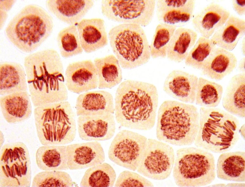
All cells of multicellular organisms originate from a single cell that divides to form the entire organism. For example, a human individual originates from a fertilised egg cell.
Cell differentiation is controlled by various signalling molecules, such as hormones secreted by endocrine glands, tissue hormones, growth factors and neurotransmitters. These are discussed in later chapters of this book. The mechanisms of differentiation are examined in more detail in the chapter on fetal development.
Stem cells are cells that are capable of differentiating into different cell types.
Embryonic cells are totipotent, meaning they can differentiate into any type of cell. As division progresses, they become pluripotent, capable of producing all the different cell types in the body, but not the placenta. However, as cell division progresses, the differentiation capacity of embryonic cells decreases. The cells of a one-week-old embryo can only differentiate into certain specific cell types, so they are called multipotent cells.
Adult stem cells can only differentiate into one type of cell. These stem cells are unipotent. The body needs these stem cells, especially when damaged or dead cells in tissue need to be replaced with new ones. However, with ageing, adult stem cells lose their ability to regenerate tissue.
In general, differentiated cells, such as nerve cells, are unable to transform back to stem cells.
Thanks to biotechnology, it is now possible to genetically engineer stem cells. These cells are known as induced pluripotent stem (iPS) cells. Researchers can now guide cells isolated from adult human tissue to revert to a state resembling that of embryonic stem cells.
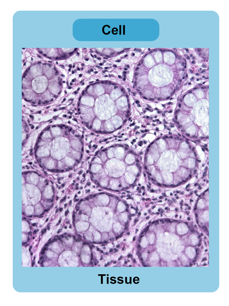
There are over 200 different types of cell in the human body. These cells form smaller and larger entities called tissues[term: tissue – A group of cells that are similar in structure and function. For example, muscle tissue.]. In human tissue, cells are held together by a substance known as the extracellular matrix[term: extracellular matrix – A substance that connects cells to form tissues. It consists of proteins and associated carbohydrate chains. The cell membrane often connects it to cells. Examples of proteins in the extracellular matrix include collagen and elastin.]. This substance is made up of molecules such as collagen (a fibrous protein), long sugar chains, and associated proteins. It also contains interstitial fluid, which is important for the exchange of substances between tissues and cells. Interstitial fluid can account for up to 20% of a person's weight. Its chemical composition remains similar to that of blood plasma because substances are constantly exchanged between the interstitial fluid and the capillaries.
From cells to tissues
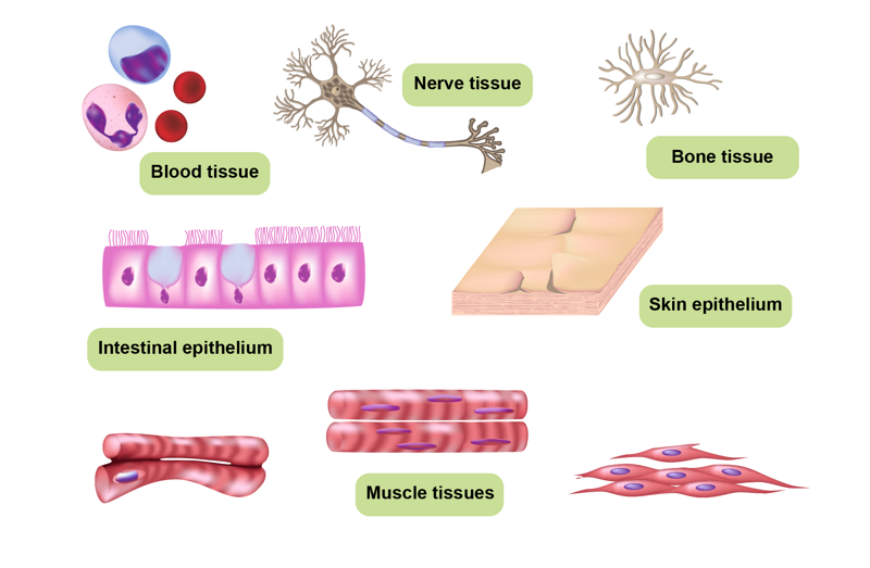
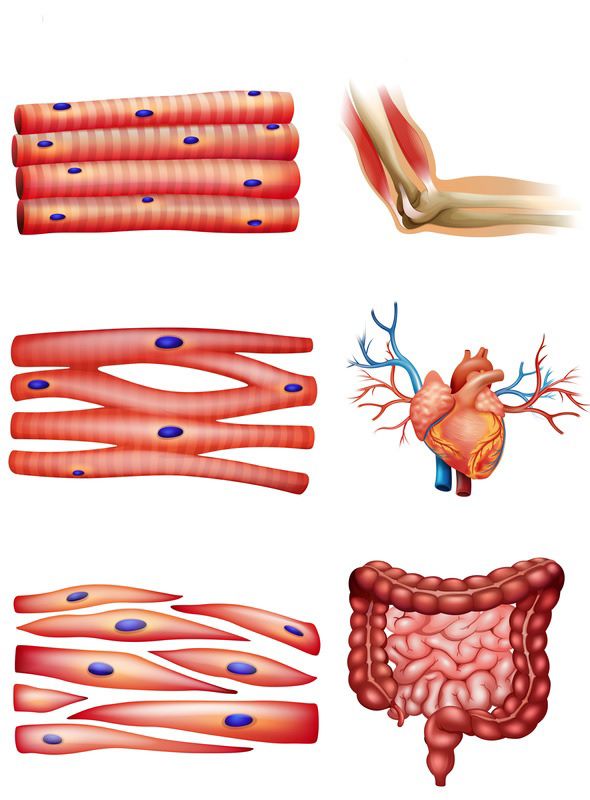
In the human body, cells that are similar in structure and function join together to form tissues. The four main types of tissue found in the human body are epithelial tissue, connective tissue, muscle tissue and nerve tissue.
Different types of tissue form organs that perform specific functions within the body. For example, the human intestines consist of intestinal epithelial tissue, two layers of smooth muscle tissue, and nerve tissue. Organs that perform the same function together form an organ system. For example, the digestive system consists of the teeth and salivary glands, the stomach, the small and large intestines, and many other parts. Other organ systems include the circulatory, reproductive and respiratory systems.
Different organs of the body also work together. For example, the digestive system breaks down and absorbs sugars into the bloodstream, which the circulatory system transports to the cells.
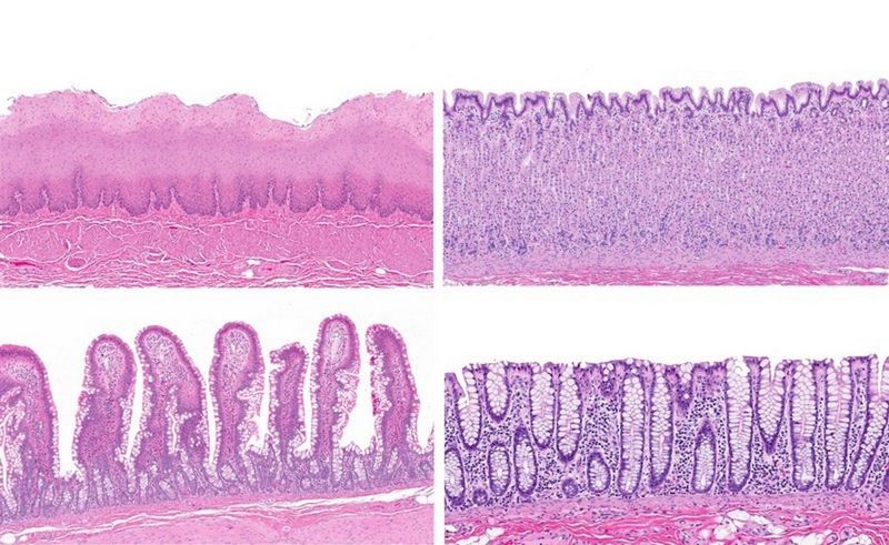
Epithelial tissue
Epithelial tissue[term: epithelium – A type of protective tissue. The epithelium separates the body from its environment. It also protects and insulates the individual. For example, the skin, the inner surface of the intestine, and the glands are epithelium.] acts as an interface between the body and its environment. It covers surfaces of the body that are in contact with the outside world, such as the skin and intestines, as well as some internal organs, including blood and lymph vessels. It also forms sacs that protect internal organs.
Epithelial tissue protects organs and organ systems, and regulates the absorption and removal of substances. It also secretes substances and acts as a sensory organ. For example, the skin provides mechanical protection for the body, prevents microbes from entering it, and reduces the evaporation of fluids from the body. Multilayered epithelium is found in organs such as the kidneys. Ciliated epithelium lines the inner surface of the respiratory tract, helping to remove dust and viruses.
Connective and supporting tissue
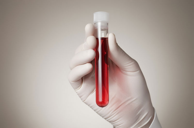
The function of connective and supporting tissue[term: connective tissue – A type of tissue that supports the body and connects organs to each other. Examples include bone, cartilage, blood, and fat.] is to connect organs to each other, as well as to support and protect the body and its organs. This tissue is connected by an extracellular matrix that is secreted around the cells. It provides the tissue with its supporting and connecting properties. Connective and supporting tissue also includes adipose tissue, cartilage, bone tissue, blood and lymphatic tissue.
Muscle tissue
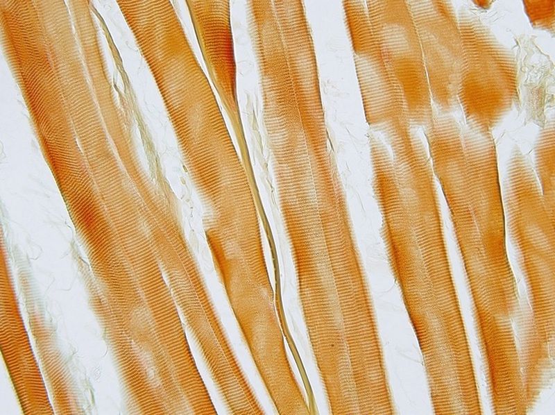
Muscle tissue[term: muscle tissue – Muscle tissue enables animals to move. It consists of contractile muscle cells that are tightly packed together. It is divided into skeletal muscle tissue, cardiac muscle tissue, and smooth muscle tissue.] consists of cells that can contract. It is responsible for producing movement in the body, for example in the intestines and skeletal muscles. There are three types of muscle tissue: striated, cardiac and smooth.
Nerve tissue
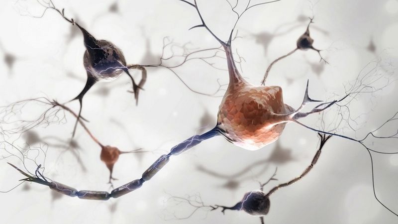
The function of nerve tissue[term: nerve tissue – A type of tissue responsible for communication and regulating other bodily functions. It consists of nerve cells (neurons) and support cells (glial cells).] is to receive, transmit and process information obtained from the environment. It consists of nerve cells, which are called neurons, and support cells, which are called glial cells.
The body needs all types of tissue in order to function. The circulatory system, for example, contains all of the aforementioned tissue types. Epithelial tissue surrounds blood vessels, while the blood flowing inside them is made up of connective and supportive tissue. While the heart is made up of muscle tissue, nerve tissue is also required to regulate its function and that of the blood vessels.
Cancer is uncontrolled cell division
Mutations occur constantly in human body cells. However, only mutations that occur in germ cells can be passed on to offspring.
Mutations can occur as errors during DNA replication or spontaneously. In terms of ageing, the key factor is the efficiency with which DNA repair enzymes function. In older individuals, these enzymes often do not function as efficiently.
Mutagens are substances or forms of radiation that cause mutations in DNA. Mutations occur naturally, and the human body has cellular mechanisms that repair them. However, not all mutations can be repaired by repair enzymes. In certain situations, cells undergo programmed cell death (apoptosis). Cells are most sensitive to mutations during cell division. For this reason, the developing fetus must be protected from mutagens, such as certain chemicals.
Carcinogens are substances that cause cancer. Many carcinogens are also mutagens. The most well-known examples are UV radiation, X-rays and radioactive radiation, as well as various chemicals found in tobacco smoke.
A tumour arises when the body’s cells begin to grow and divide uncontrollably. Tumours can be either benign or malignant. Benign tumours stay confined to one area and are generally straightforward to manage. Malignant tumours are called cancers. They invade nearby tissues and can spread to distant sites in the body, forming metastases.
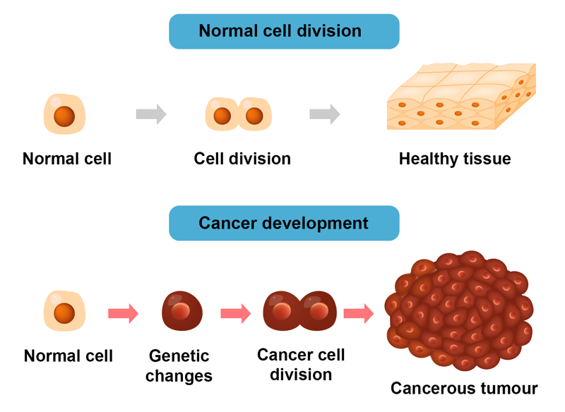
The development of cancer involves changes or mutations that occur within the cell and a reduction in the influence of surrounding cells. There are over 200 different types of cancer. The only common factor found in different cancers may be that the factors regulating the division of cancer cells cease to function.
A cancerous tumour takes over the body by increasing blood vessel growth. When blood vessels grow into the tumour, it receives the energy and nutrients it needs to grow. Blood vessels also allow cancer cells to spread to other parts of the body.
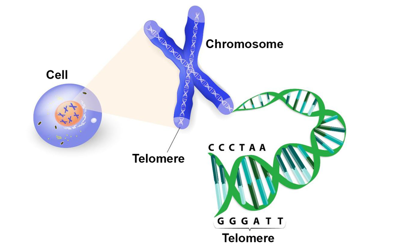
Telomeres are repetitive DNA sequences that protect the ends of chromosomes. Cancer cells produce telomerase, an enzyme that keeps telomeres long enough to prevent cells from becoming unable to divide. Therefore, in cancer cells, the shortening of telomeres does not limit cell division like it does in normal cells. This property of telomerase has also been exploited in cell cultures. The oldest cell line in use is HeLa, which was established in 1951 from the cervical cancer cells of Henrietta Lacks.
Cancer is caused by mutations in genes that regulate cell division. These include proto-oncogenes which, when mutated, become oncogenes that promote growth, and tumour suppressor genes which, when inactivated, lose their ability to control growth. Cancer genes are often large developmental genes that regulate cell growth and differentiation. Due to their genetic background, some individuals may be predisposed to cancer. For instance, some gene variants which increase breast cancer risk may have conferred evolutionary benefits by regulating telomeres and keeping egg cells in better condition. However, these gene variants also facilitate tumor development.
It can take a long time for the necessary mutations to accumulate, which is why older people are more likely to develop cancer than younger people. In addition, the human body often recognises cancer cells, whose cell membranes carry antigens produced by cancer genes. As the individual ages, their immune system weakens, which also increases the risk of developing cancer; essentially, the weakened immune system lets cancer "slip through its cracks". It has also been suggested that epigenetic factors — the regulation of gene activity during individual development — contribute to the development of cancer. In many cancers, the cells responsible for tumour growth are cells that have begun to function like undifferentiated stem cells for one reason or another.
Mutagens can accelerate mutations. There are also substances that are carcinogens but not mutagens. Cancer is generally not contageous. The development of cancer can be influenced by pathogens such as Helicobacter pylori, which causes stomach cancer. We ingest carcinogens through our diet, for example from red meat, particularly when it is processed or grilled. Obesity can also increase the risk of developing cancer.
If detected early, cancerous tumours can be surgically removed if they are made up of tissue that can be cut away. However, if the cancer has already spread to other parts of the body (metastasised), chemotherapy is needed to prevent or slow down tumour growth. Another method of treatment is radiation therapy. It works by destroying cells during the division phase, when rapidly dividing cancer cells are more vulnerable than other cells in the body.
Common cancer types
Cancer | Information |
Breast cancer | The most common cancer in women. |
Prostate cancer | The most common cancer in men. |
Lung cancer | Common in both men and women. |
Skin cancer | Has become more common in all age groups. There are many types of skin cancer, such as melanoma. |
Leukemia | A blood cancer caused by immature white blood cells transforming into cancer cells. Rather than producing a single tumour, the cancer cells circulate in the blood and bone marrow. |
Summary
- The four types of stem cells are totipotent, pluripotent, multipotent, and unipotent stem cells.
- Human cells form tissues, tissues form organs, and organs form organ systems.
- Tissues include epithelial tissue, connective and supporting tissue, muscle tissue, and nerve tissue.
- Carginogens are substances that cause malignant tumors, or cancers. Most of these substances are also mutagens.
- The best-known mutagens include UV radiation, X-rays, and radioactive radiation, as well as various chemical substances, such as tobacco smoke.
- All types of cancer are caused by the failure of the factors that regulate the division of cancer cells.